Medical Basis
Physicians, you may find our combination therapy useful for selected
patients with aggressive malignancies for whom standard treatment offers little potential for disease control or better.
Our initial human experience with this novel technique has yielded extremely promising results in several of the diagnoses
which challenge us the most and the outcomes are now improving even further as we define which patients may be able to benefit
from additional incorporation of other agents in precisely synchronized and monitored fashion.
This outpatient technique is an embodiment of combination immunotherapy
which was devised and implemented nearly fifteen years ahead of the current research rush in that direction. It has
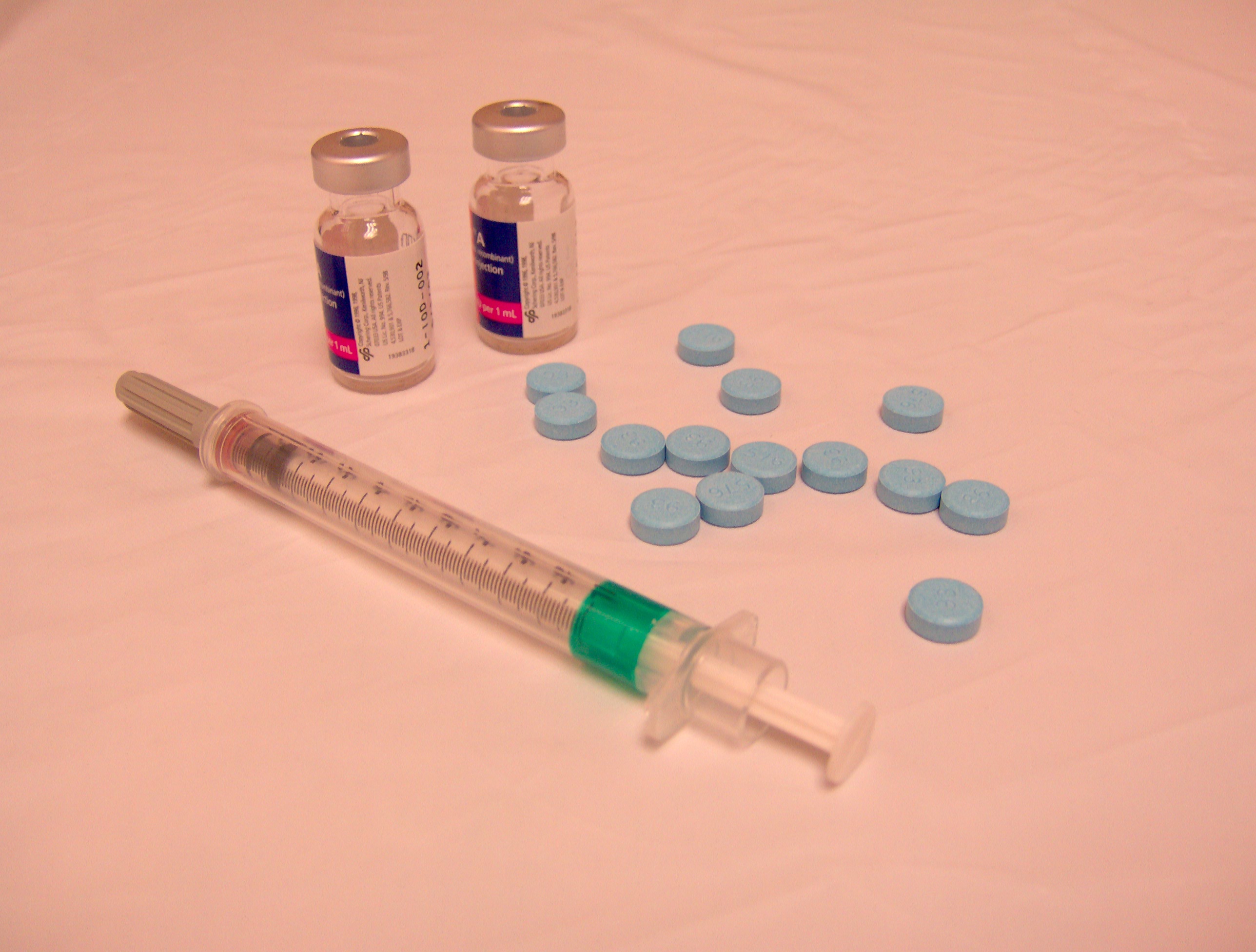
at its heart a combination of low-dose interferon alfa-2b and lovastatin. The latter has long been known
to have significant antineoplastic properties in vitro and has started to receive a good deal of renewed research attention
in the last few years, especially with emerging findings of its chemopreventive benefit and synergistic enhancement of a number
of cytotoxic regimens. Our experience over the last fifteen years has been that it provides benefit to
certain patients at very safe and tolerable doses, with surprising efficacy when administered in combination with interferon.
For most of our patients cimetidine is also used, and one or more agents from capecitabine, temozolomide, and hydroxychloroquine
may be added when appropriate. There are also several diagnoses in which we have found our therapy to be quite beneficial
but even more so if a standard cytotoxic regimen (with total dosing possibly reduced to approximately 60-70% of typical) is
provided concurrently. Thus we have a growing number of patients who maintain active treatment with us and their primary
oncologist.
This
treatment is currently most appropriate for qualifying patients with:
Pancreatic
adenocarcinoma and cholangiocarcinoma
Melanoma
Colon cancer
and mucinous adenocarcinoma ("PMP")
Mesothelioma
Rhabdomyosarcoma, MFH, osteosarcoma, chondrosarcoma,
DSRCT
We
will encourage any of your patients who may choose to enter this program to continue to see you regularly as well. We strongly
prefer that every patient maintain an active doctor-patient relationship with an independent oncologist and other specialists
where appropriate. Referring physicians and other treating physicians will be kept informed of progress on a regular
basis. You can be assured that the good work you're doing will be respected and appreciated here, not criticized.
Please peruse the entirety of our web site at your leisure, and do
not hesitate to let us know of further information we can provide. Thanks for visiting!
Criteria for
treatment
The best candidates for treatment are those meeting the following criteria.
1. No history
of cerebral metastases. Patients with cerebral mets are accepted if appropriate therapy for those lesions continues
simultaneously.
2. Ability to
absorb oral medications (or via feeding tube).
3. ECOG performance status of 0, 1, or 2.
4. Hepatic
enzymes not greater than twice upper limit of normal reference range.
5. No history of cirrhosis or other non-cancer-related
hepatic pathology (with the exception of hepatitis B and C which are potentially compatible with treatment and may also benefit
from it).
6. Not pregnant.
We also recommend the following for the diagnoses listed.
Pancreatic cancer: Surgical resection to the extent possible. Surgical re-routing of bile duct, or functioning
stent in place. No biliary stasis. Serum bilirubin values not greater than 1.5 times upper limit of normal reference
range. We prefer our treatment to run concurrently with gemcitabine but this is not a requirement.
Melanoma:
Surgical resection to the extent possible, with sentinel lymph node dissection recommended. PET scan strongly recommended
at start of treatment.
Colon cancer: Surgical resection to the extent possible.
Renal cell carcinoma: Surgical resection to the extent possible.
Mesothelioma: Surgical resection
to the extent possible. Entry at any time after diagnosis; preferably, our treatment to run concurrently with first-line
treatment (pemetrexed with or without additional agent) if not already administered.
Malignant fibrous histiocytoma,
osteosarcoma, chondrosarcoma, DSRCT: Surgical resection to the extent possible. Prior failure (disease progression
or other failure to respond) to first-line therapy, or treatment to run concurrently with first-line treatment if not already
administered.
Non-qualifying patients
Patients who do not meet the criteria above may be considered for
compassionate treatment with the same regimen if they are experiencing disease progression with a poor prognosis after completing
indicated therapies. If so requested by a patient, this determination will be made on a case-by-case basis.
Theoretical basis
and possible mechanisms
Our primary goal is providing the greatest possible benefit to each of our patients
while also collecting relevant data on the use of this new treatment method. As such, our clinical work follows
the hypothesis that the combination of statin agents and interferons is effective in the treatment of certain aggressive
malignancies in a highly synergistic fashion. As in vivo evidence of statins' cancer efficacy continues
to accumulate, a rich background of in vitro data already portrays five or six interrelated effects of lovastatin and certain
of its analogues on malignant cells.
The repeated finding that lovastatin has substantial activity against malignant cell lines while posing little known
threat to normal ones suggests that the malignant cells employ an aberrant process or feature which can be targeted.
Such is the first mechanism: many malignant cells are unusually dependent on de novo synthesis of
cholesterol, especially of LDL composition, for growth and membrane integrity. The dependence on the mevalonate
pathway for the necessary supply explains the inability of the cells to grow and proliferate when this path is disabled.
Given sufficient continuous concentration of the drug over a period of time, apoptosis is induced in a number of malignant
cell lines. It is postulated that fragility of the membrane may leave the malignant cell susceptible to
damage and death on its own or after exposure to other agents. Therefore the effects may be cytostatic
or cytocidal depending on dose and duration of exposure and characteristics of the individual cancer cell line.
The second observation is probably the most crucial and may
increasingly be proven the root mechanism for several other anti-neoplastic effects. Lovastatin and its
analogues interfere with myriad post-translational protein processing reactions in the malignant cell, thus preventing critical
proteins from being expressed on the membrane or in the nucleus. Striking at such basic processes no doubt
has the capacity to produce cytostasis or cellular death in many forms. It is not understood why this doesn’t
cause more discernible problems in normal cells, but it may be responsible for some of the few side effects which are occasionally
seen and we suspect this may be demonstrated with more time and research.
The third effect was recognized shortly after the discovery of compactin and lovastatin. Cancer
cells are prevented from completing mitosis in the presence of sufficient concentration of the hmg-CoA reductase inhibitors
(HRIs), specifically in the G0 or G1 phases and between G2 and M. This
cycle arrest is cytostatic only and in most cases can be reversed when the drug is withdrawn or mevalonate is added.
Future research may well reveal this to be due to specific defective proteins as mentioned above.
A fourth occurrence is closely related to the second and third phenomena
discussed. When a cell does initiate mitosis, lovastatin interferes with DNA synthesis
and more specifically prevents proper formation of daughter nuclei after DNA replication. This may leave
the nascent DNA exposed and “marked for destruction” in a cascade resulting in apoptosis and ultimately necrosis.
A fifth area of attack has been better characterized recently and
carries profound implications. Lovastatin has been shown both in vitro and in animal models to interfere
significantly with the metastatic activity of a number of malignancies, usually by preventing adherence to and migration across
endothelial surfaces. This may be due to inhibition of the cell's ability to express certain membrane proteins necessary
for this metastatic sequence.
Lovastatin has also been shown to impede tumor-induced angiogenesis in a melanoma model in mice, making it comparable
in this respect to many other drugs now under intense study. This action could be helpful in larger tumor
masses but we do not believe the anti-neovascular effect, of itself, has potential for eradicating disease or preventing metastasis.
It seems more likely that other mechanisms are chiefly responsible for the observed benefits, coincident with but not
directly due to impairment of angiogenesis. These various agents may eventually prove to have much in common
with the anti-neoplastic mechanisms of the HRIs.
Until recently the subject of interferon in melanoma and other malignancies has been a conundrum.
There is no doubt that it has been of value to some individuals but the data are inconsistent among various experiences.
Overall, its value has been marginal even when statistically significant: meta-analyses in melanoma confirm
that, used alone, it confers a few extra months until relapse to about 15% of patients and no overall survival increase at
all. Since lovastatin administration in vitro can diminish natural killer (NK) cell cytotoxicity and interferon
gamma production, Dr. Cantrell hypothesized in 2000 that concurrent exogenous interferon administration might be
able to overcome any such down-regulation and in fact to enhance activity of the same cells in reverse fashion.
(This was admittedly a "shot in the dark" as no prior work suggested such a relationship.) It is now
apparent that the activity of interferon against malignant cells is primarily due to an enhanced effect of NK cells
and/or other components of the native immune system. It is further postulated that such activity is made more effective
by impairment of the integrity of those cells due to the actions of lovastatin as described above, thus possibly providing
the needed signal for an immune reaction to suppress or even destroy cancerous cells. The clinical and in vitro
data to support or negate that hypothesis will emerge over the coming years, but the patient outcomes are already supporting
the efficacy of the technique in a number of cancer types.